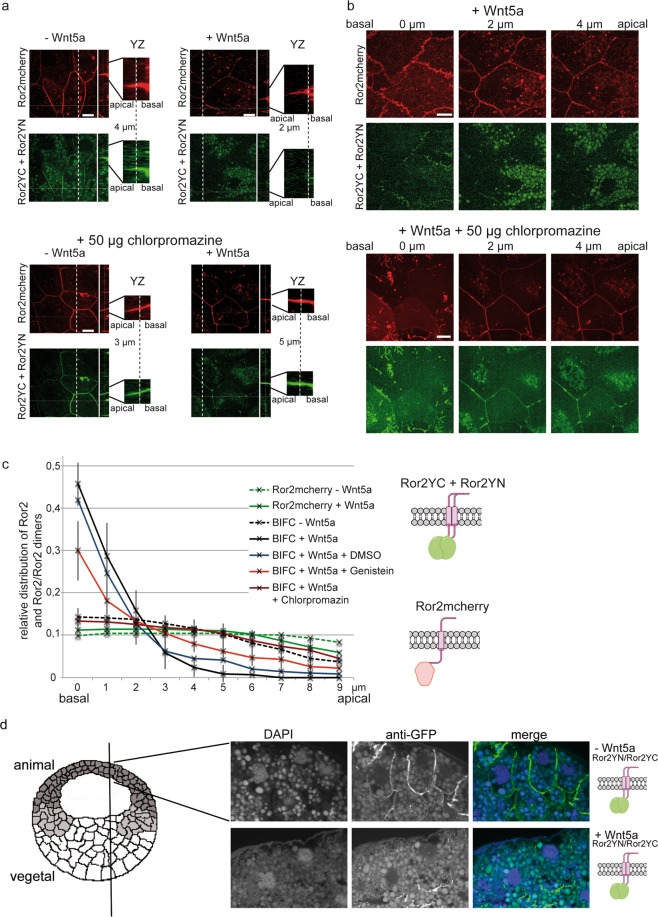
Figure 5.
Wnt5a induces disappearance of Ror2/Ror2 dimers at the apical membrane. (a) Animal cap explants were analyzed for the localization of total Ror2 (Ror2mCherry, red channel) and Ror2 homodimers (splitYFP signal, green channel) in the absence (−Wnt5a) and presence (+Wnt5a) of co-injected Wnt5a and in the presence of 50 µg chlorpromazine. Ten optical sections of 1 µm thickness allowed us to visualize localization of the signals in the z-dimension. The white bar in the close-ups of the yz-plane marks the z-coordinate shown in the xy-images. (b) xy-planes at three different z-values illustrate the exclusively basal and basolateral localization of Ror2/Ror2 dimers in Wnt5a co-injected explants. (c) To quantify the z-distribution of the fluorescence signals, at least 11 explants (each with 4 to 6 different cells) derived from 3 independent experiments were analyzed. Individual fluorescence spots were selected in the maximum intensity projections, and the images were analyzed section by section for visibility of these spots. Shown is the fraction of fluorescence spots found in the xy-images as a function of the z-coordinate from basal (0 µm) to apical (9 µm). These data were calculated for each cell by dividing the number of fluorescence spots of a particular xy-plane by the number of spots in all planes. Shown are mean values and standard deviations from at least 11 explants. It is apparent that the BiFC signal (Ror2 homodimers) gets shifted to the basal side in the presence of Wnt5a; chlorpromazine and genistein revert this effect. (d) Paraffine sections of early gastrula stages revealed that Ror2 homodimers are located at the entire lateral site of animal cap cells in the absence of Wnt5a. In the presence of Wnt5a, however, the dimers are found exclusively at the basal side.

