Figure 9.
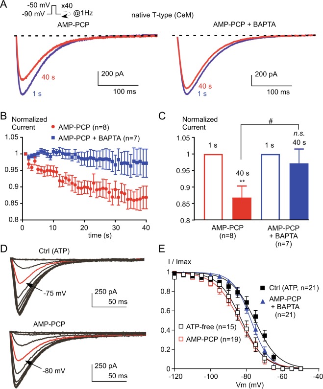
The native T-type current from neurons of rat central medial (CeM) nucleus is regulated by an interplay between a rise in intracellular calcium and a phosphorylation mechanism. (A) Typical T-type current elicited by stimulation at −30 mV applied at a frequency of 1 Hz from a holding potential of −90 mV obtained from CeM neurons dialyzed with either AMP-PCP or AMP-PCP combined with BAPTA. (B) Normalized current amplitude from 1 to 40 s. (C) Average normalized current amplitude recorded at 1 s and at 40 s. Asterisks indicate significant difference in the current amplitude at 40 s as compared to the current amplitude at 1 s. The hash signs indicate statistical difference between AMP-PCP and AMP-PCP + BAPTA. (D) Representative T-type current recorded at −50 mV from holding potentials ranging from −120 to −50 mV (5 mV increment, 3.6 s duration). CeM neurons were dialyzed with either ATP (control condition) or AMP-PCP. The red trace indicates the half-inactivation potential. (E) Average steady-state inactivation curves obtained from experiments presented in (D) for neurons dialyzed with ATP (control), AMP-PCP, AMP-PCP combined with BAPTA, and in absence of ATP (ATP-free).

