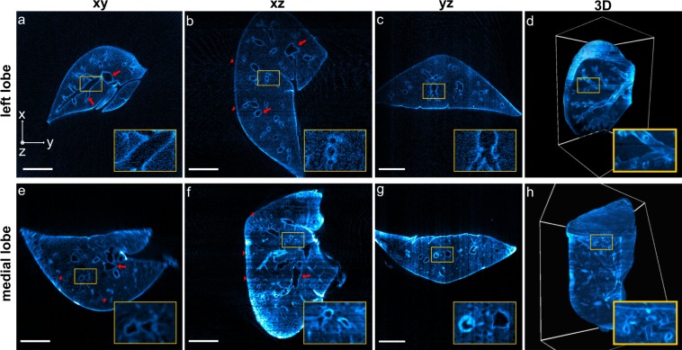
Figure 3.
OptiJ reconstructions of murine lungs. Reconstructions of a left lobe labelled with anti surfactant C – Alexa Fluor 488 (a–d) and a medial lobe labelled with anti TTF1 – Alexa Fluor 488 (e–h) from 512 eOPT projections, displayed in xy, xz, and yz orthogonal views (left three columns), as well as rendered in 3D (right-most column). (a–d) The red arrows and the insets indicate the primary airways visualized in the orthogonal cross-sections. The 3D rendering in panel d displays a clipping plane through the lung, highlighting secondary and tertiary bronchi in the inset. (e–h) The red arrows indicate a set of main airways (secondary and tertiary bronchi) in the medial lobe, and red arrowheads indicate high-order airways inside or close to the parenchyma. Small airways close to the primary bronchi are highlighted in the insets on panels e and f. The 3D rendering in panel h with a clipping plane on one of the lobe faces shows a thick meshwork of higher order airways (quaternary bronchi and bronchioles). Interactive 3D renderings are available in our online repository.

