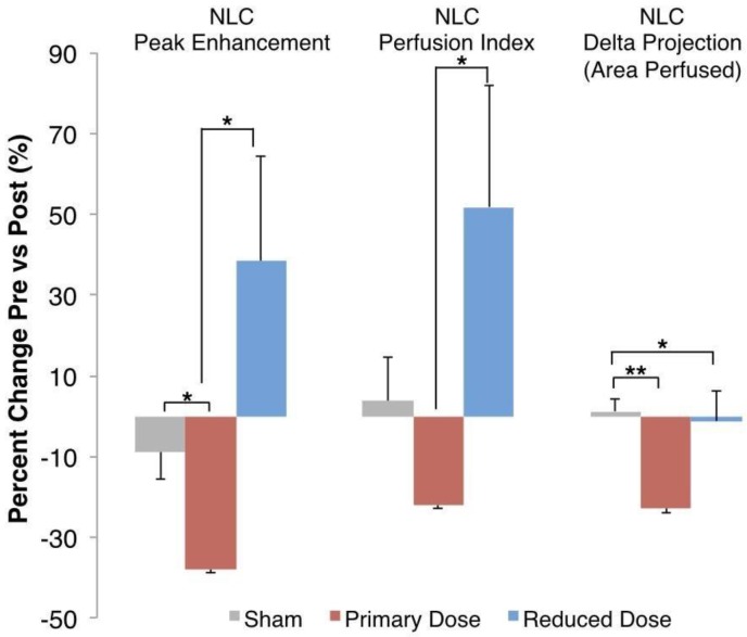
Figure 3.
Percent Change in Perfusion Parameters. Nonlinear contrast ultrasound showed a decrease in blood volume perfused of 37.9% ± 10.05% in each tumor before and after primary-dose AVUS, while perfusion only decreased 8.8% ± 6.8% in the sham arm (p = 0.02). Perfusion per tumor in the reduced-dose arm increased compared to primary-dose AVUS (p = 0.02) but was not significant versus the sham arm (p = 0.11). Perfusion index showed a similar pattern, but did not show significant differences versus sham treatment. Delta projection, quantifying the tumor area perfused throughout the scan, showed a constant perfusion in sham (increase of 1.2% ± 3.2%) and a decreased area of the tumor receiving perfusion with contrast agent after primary-dose AVUS (decrease of 22.9% ± 7.3%, p = 0.005).

