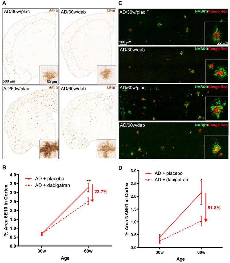
Figure 5. Long-term anticoagulation with dabigatran ameliorated amyloid burden in AD mice.

Aβ brain pathology was analyzed in TgCRND8 and WT mice treated with dabigatran/placebo. (A) Aβ immunohistochemistry (6E10) showed widespread amyloid pathology throughout the brains of 30- and 60-week-old TgCRND8 AD mice (insets show high magnification of cortical amyloid plaques). Sections and quantified cortical areas are outlined for clarity. (B) 6E10-positive cortical area was decreased by 23.7% in 60-week-old dabigatran-treated TgCRND8 mice compared to placebo. (C) Aβ oligomer immunofluorescence analysis (NAB61, green) showed staining around fibrillar amyloid plaques (Congo Red, red) in the brains of TgCRND8 mice (see insets for high magnification). Confocal tile-scan Z-stacks were acquired over the parietal cortex and quantification of the NAB61-positive area showed dabigatran treatment decreased by 51.8% the amount of oligomeric Aβ in the cortex of 60-week-old AD mice (D). Graphs show mean±SEM. Effect of treatment (p=0.0089 for 6E10 & p=0.0083 for NAB61) and age (p˂0.0001 for both) are significant. *p≤0.05, **p≤0.01 AD/60w/placebo vs. AD/60w/dabigatran. n=5-11 mice/group. Abbreviations as in Figure 1.
