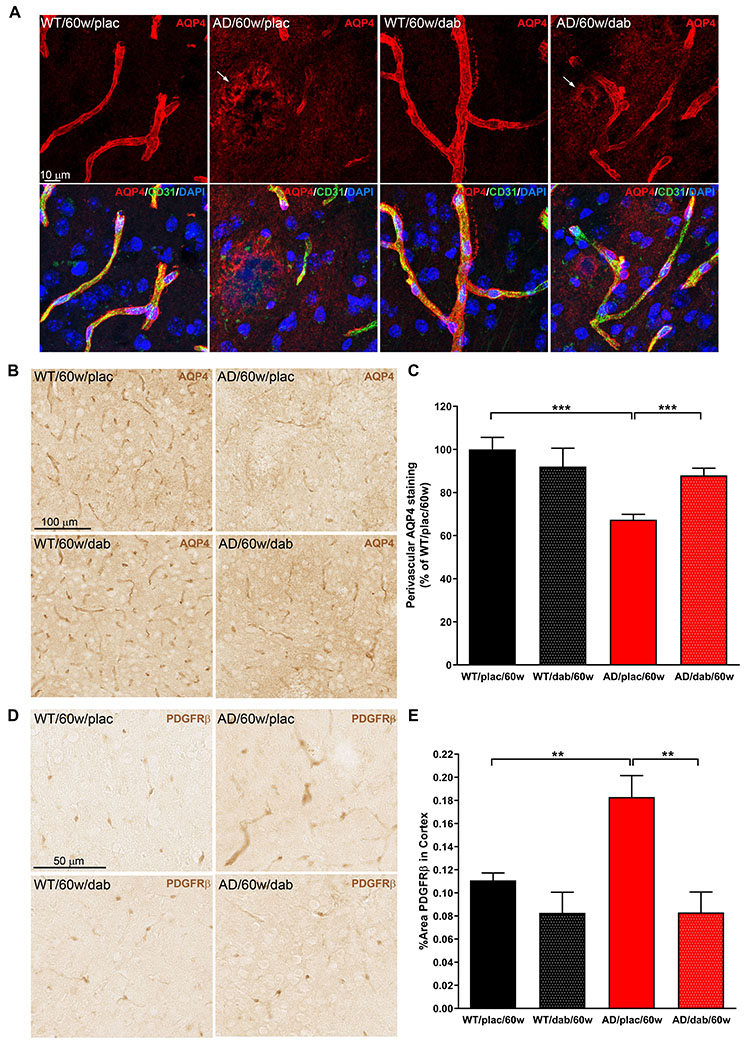
Figure 7. Dabigatran treatment preserved AQP4 expression and pericyte morphology at the BBB.

Immunostaining was performed on brain slices from 60-week-old TgCRND8 AD and WT mice treated with dabigatran/placebo to detect expression and localization of AQP4 and PDGFRβ. (A) Double immunofluorescence showed that AQP4 expression (red) was found around blood vessels (CD31, green) in the astrocytic endfeet of WT mice. However, AQP4 staining shifted from its perivascular location to neuropil areas surrounding amyloid plaques in AD mice (arrows). (B) Immunohistochemical analysis was performed to segment and quantify the amount of perivascular AQP4 in all experimental groups (C). AD mice treated long-term with dabigatran presented significantly more perivascular AQP4 staining than placebo-treated AD mice. (D) PDGFRβ immunohistochemistry demonstrated hypertrophic pericytic processes outlining the capillaries of TgCRND8 AD mice. (E) Quantification showed a robust increase of PDGFRβ-positive area in the cortex of 60-week-old AD mice compared to WT mice. This abnormal pericyte staining was normalized by dabigatran treatment. Graphs show mean±SEM. **p≤0.01; ***p≤0.001. n=5-6 mice/group. AQP4=Aquaporin 4; PDGFRβ=Platelet-Derived Growth Factor Receptor-β. Other abbreviations as in Figure 1.
