
Fig. 3. Hepatocyte Notch activation exacerbates NASH and fibrosis.
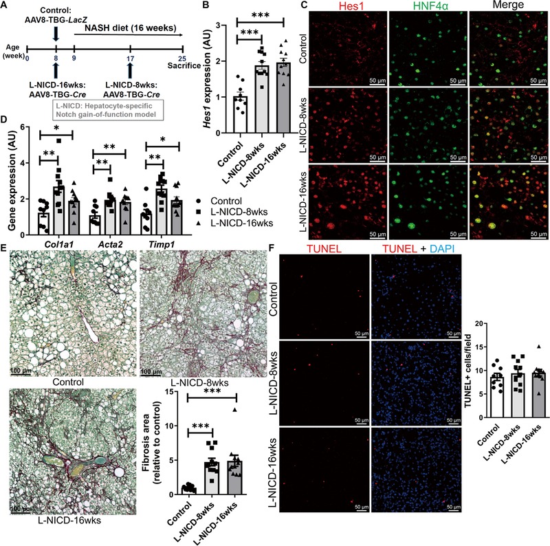
(A) RosaNICD mice (8 weeks old) were transduced with AAV8-TBG-LacZ (control) or AAV8-TBG-Cre to generate hepatocyte-specific Notch gain-of-function (L-NICD-16wks) mice before 16 weeks of NASH diet feeding or halfway through NASH diet feeding (L-NICD-8wks) (n = 9 to 11 per group). (B) Hes1 expression and (C) representative images of Hes1 (red) and HFN4α (green) staining, (D) expression of fibrogenic genes, (E) collagen staining and quantification, and (F) TUNEL (red) staining in livers from control and L-NICD mice. *P < 0.05, **P < 0.01, and ***P < 0.001 as compared to Cre− control mice by one-way ANOVA, followed by post hoc t tests (three groups). All data are shown as the means ± SEM.