Figure 1.

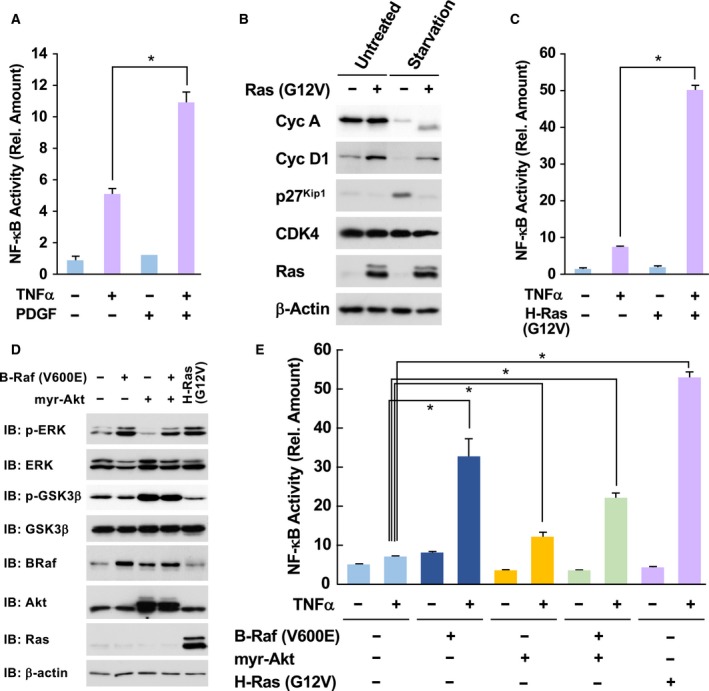
Oncogenic Ras mutants accelerate TNFα‐induced NF‐κB activation. (A) KF‐8 cells were cultured in DMEM without FBS for 24 h. Then, cells were stimulated with 10 μg·mL−1 TNFα with/without costimulation with PDGF (10 μg·mL−1) for 16 h. Cells were lysed, and the activity of expressed luciferase levels was measured as NF‐κB activation. (B) KF‐8 cells were infected with control retroviruses or retroviruses harboring H‐Ras (G12V). After puromycin selection, the expression of cell cycle‐related proteins, such as cyclin A (CycA), cyclin D1 (CycD1), Cdc2, CDK4, and H‐Ras, was detected by immunoblotting analysis. β‐actin was loading control. (C) Infected KF‐8 cells as shown in (B) were cultured in DMEM without FBS for 24 h. Cells were stimulated with 10 μg·mL−1 TNFα, and then, the activity of luciferase was analyzed. (D) KF‐8 cells were infected with control retroviruses or retroviruses harboring indicated cDNA. Then, downstream signals such as the phosphorylation of ERK and GSK3β were analyzed. (E) Using infected cells shown in (D), the luciferase assay was performed. In graphs, error bars indicate SD (n = 3), and the results of calculations of independent t‐tests are shown (*P < 0.005).
