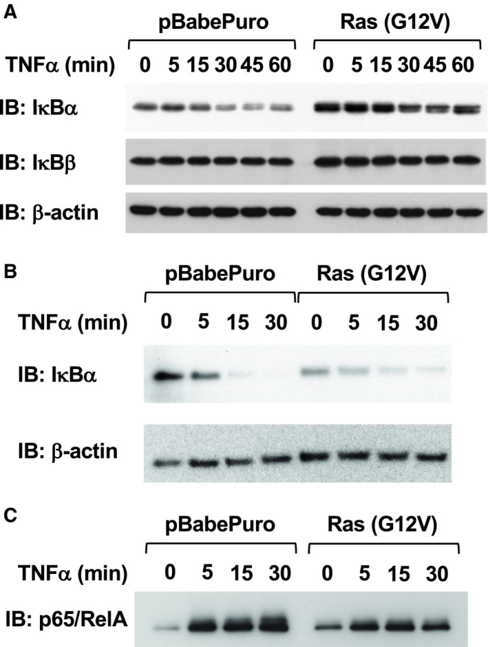
Figure 2.

Effect of H‐Ras (G12V) on TNFα‐induced signaling pathways. (A) Parental NIH‐3T3 cells were infected with control retroviruses or retroviruses harboring H‐Ras (G12V). After puromycin selection, cells were stimulated with 10 μg·mL−1 TNFα for the indicated periods. Then, the cells were lysed, and the degradation of IκBα and IκBβ was analyzed by immunoblotting analysis. (B) Cells were treated with 25 μg·mL−1 CHX for 30 min, and then, the cells were stimulated with 10 μg·mL−1 TNFα for the indicated periods. Cell lysates were prepared, and the degradation of IκBα was analyzed. (C) From the infected NIH‐3T3 cells shown in (A), nuclear extracts were prepared. The nuclear extracts were incubated with a biotin‐labeled DNA probe harboring κB‐responsive elements, and then, DNA·protein complexes were captured using streptavidin‐conjugated agarose. The captured proteins were eluted with sample buffer. Using these samples, the nuclear localization and DNA binding activity of NF‐κB were evaluated by immunoblotting analysis for p65/RelA.
