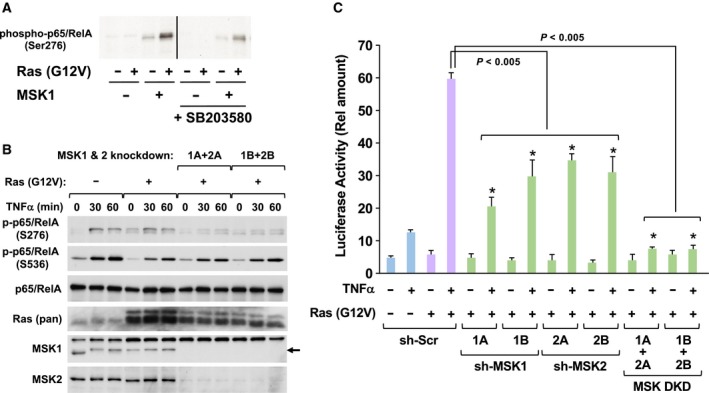
Figure 5.

MSK1/2 contributes to the oncogenic transcriptional activation of NF‐κB. (A) HEK293T cells were transfected with the indicated combinations of plasmids harboring FLAG‐MSK1 and H‐Ras (G12V). The cells were cultured in FBS‐free DMEM for 24 h, and then, the cells were lysed with NP‐40 lysis buffer. FLAG‐MSK1 was purified by immunoprecipitation with M2‐agarose, then incubated with 3 μg GST‐p65/RelA and 100 μm ATP at 30 °C for 30 min. The phosphorylation of p65/RelA at Ser276 was detected by immunoblotting analysis using an antibody against phosphorylated p65/RelA (S276). (B) NIH‐3T3 cells infected with indicated retroviruses were stimulated with 10 ng·mL−1 TNFα for indicated periods, and then, their cell lysates were prepared as described in Materials and Methods. The phosphorylation of p65/RelA at Ser‐276 and Ser‐536 was detected by immunoblot analysis using indicated antibodies, respectively. The expressions of p65/RelA and H‐Ras were also detected by immunoblot analysis using indicated antibodies. (C) KF‐8 cells were infected with the indicated combinations of retroviruses harboring H‐Ras (G12V), shRNA, sh‐MSK1, or sh‐MSK2. Then, using these infected cells, luciferase assays were performed. In graph, error bars indicate SD (n = 3), and the results of calculations of independent t‐tests are shown (*P < 0.005).
