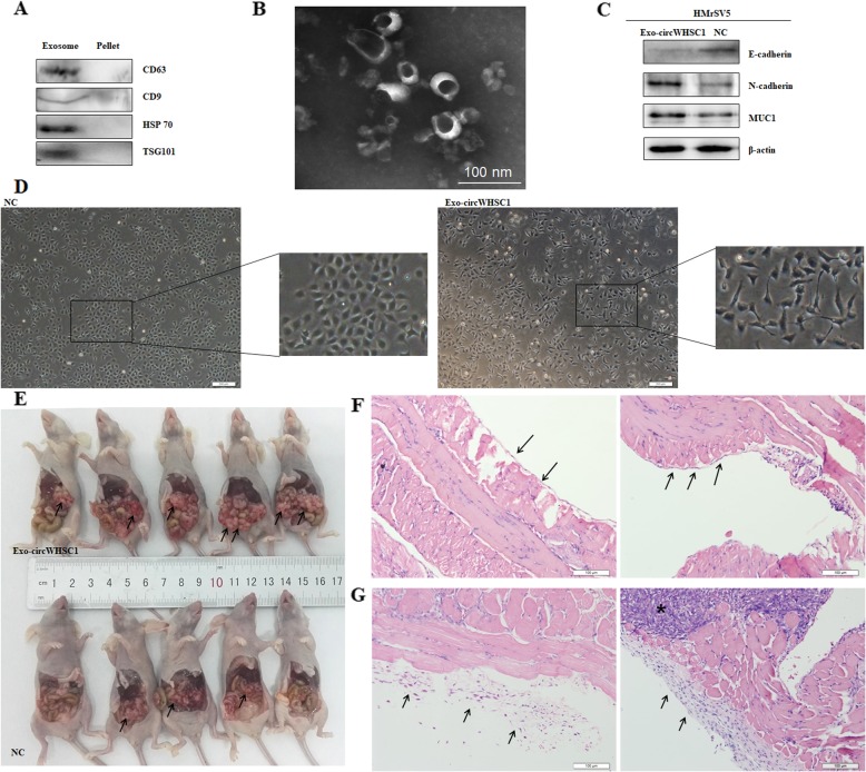
Fig. 5.
Exosomal circWHSC1 promotes peritoneal dissemination and regulates expression of MUC1 in peritoneum. Exosome specific markers, CD63, CD9, HSP70 and TSG101 were expressed in the extracted exosome pellets (a). Electron microscopy confirmed diameters of the extracted exosomes ranged from 10 to 100 nm and with cup shape appearance (b). The morphology of HMrSV5 cells incubated with exosomal circWHSC1 was converted into fibroblast-like (c). N-cadherin and MUC1 were up-regulated, while E-cadherin was down-regulated in exosome-treated HMrSV5 cells (d). After injection with circWHSC1 exosomes, the number of tumor nodules was significantly increased in abdominal cavity (e). HE staining showed that the peritoneum was covered with a monolayer of mesothelial cells with intact intercellular junctions from two different perspectives (f). After the treatment of exosomal circWHSC1, mesothelial cells arranged loosely, and the stromal layer was reactively thicken surrounding infiltration of tumor cells, and the left and right graphs represent two different perspectives (g)

