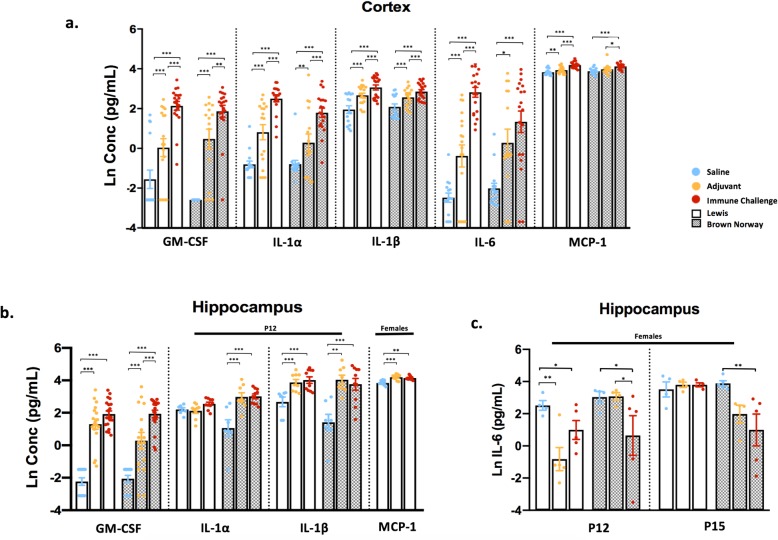
Fig. 3.
Peripheral immune stimulation broadly upregulates innate cytokine levels in the cortex and hippocampus. Cortical, hippocampal, and cerebellar lysates were collected from rats 2 or 5 days following exposure and subjected to cytokine and chemokine analysis. a Concentrations of innate cytokines in the cortex, compared between experimental conditions. Data are collapsed between sex and day due to minimal differences seen; N = 15–20 per condition. b, c Cytokine levels from hippocampal lysates; black solid bars above certain analytes specify time point or sex-specific conditions. b Levels of several innate cytokines: GM-CSF collapsed between sex and day (N = 13–20 per condition), IL-1a, IL-1B shown at P12 only and MCP-1 in females collapsed between day (N = 7–10 per condition). c Hippocampal IL-6 levels across treatment and strain in female rats; N = 4–5 per condition. Data represent mean +/− SEM, *p < 0.005, **p < 0.01, ***p < 0.001

