
Fig. 5.
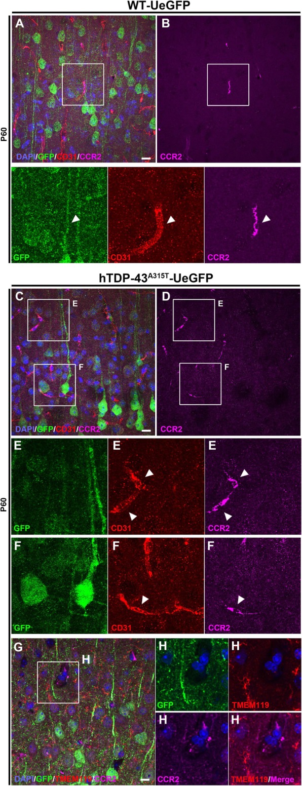
Evidence of infiltrating monocyte contribution in the motor cortex of prpTDP-43A315T-UeGFP mice. a A representative image of motor cortex of WT-UeGFP mice showing an infiltrating monocyte (CCR2+) within the blood vessel (CD31+), see arrowheads. c, d A representative image of motor cortex of prpTDP-43A315T-UeGFP mice showing higher numbers of infiltrating monocytes (CCR2+) within the blood vessel (CD31+), see arrowheads and insets enlarged bellow in E, F. g, h A representative image of motor cortex of prpTDP-43A315T-UeGFP mice showing infiltrating monocytes (CCR2+) that lack TMEM119 expression within the blood vessel. Inset is enlarged to the right. Scale bar = 20 μm