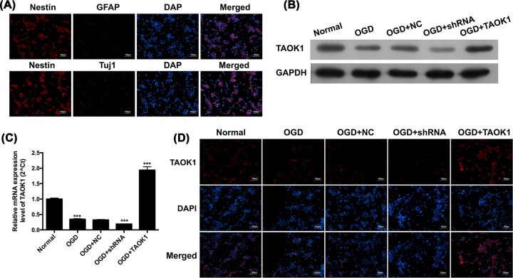
Figure 3. TAOK1 expression was significantly decreased post-OGD in neural stem cells.
(A) Identification of neural stem cells by examining the corresponding types of molecular markers, Nestin, GFAP and Tuj1 with immunofluorescent (IF) staining. (B,C) Relative protein and mRNA expression of TAOK1 of TAOK1 blocked and overexpressed neural stem cells under OGD conditions were assessed by Western blotting and qRT-PCR, respectively (***P<0.001 vs Normal group). (D) Immunofluorescent staining was performed to examine the expression of TAOK1 in TAOK1 blocked and overexpressed neural cells under OGD conditions.

