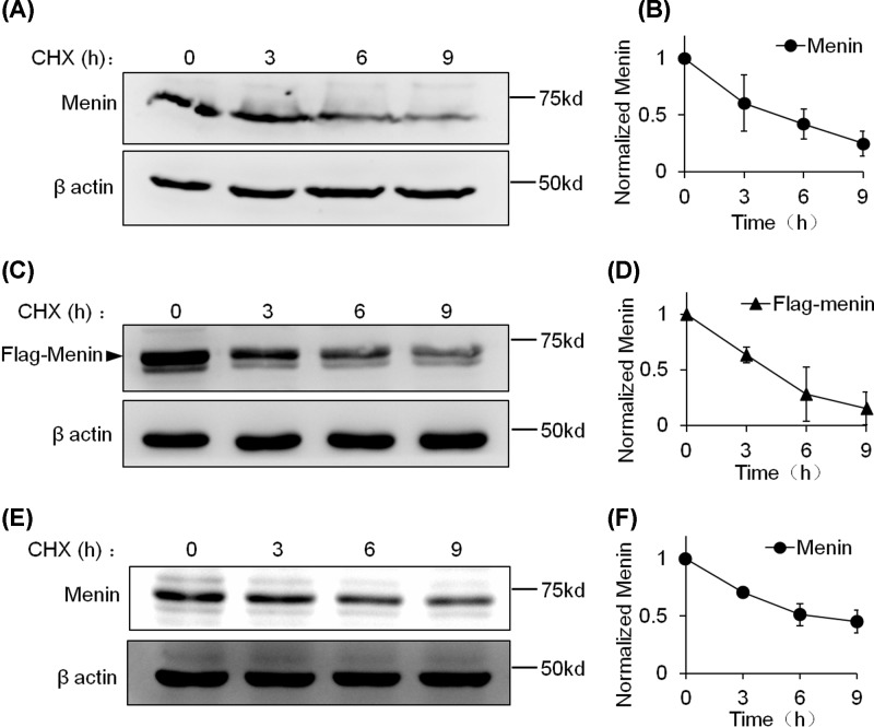
Figure 3. WT menin degrades rapidly in the presence of CHX in INS-1 cells.
(A) INS-1 cells were treated with CHX (20 μg/ml) for the indicated times and lysed for Western blot. (B) Quantitation of menin protein level from (A). Gray analysis was executed with ImageJ and menin protein levels were quantified and normalized to β-actin. (C) INS-1 cells expressing ectopic Falg-menin were treated with CHX (20 μg/ml) for the indicated times and lysed for Western blot. (D) Quantitation of the Flag-menin protein level from (C). Gray analysis was executed with ImageJ and Flag-menin protein levels were quantified and normalized to β-actin. (E) TGP-61 cells were treated with CHX (20 μg/ml) for the indicated times and lysed for Western blot. (F) Quantitation of the menin protein level from (E). Gray analysis was executed with ImageJ and menin protein levels were quantified and normalized to β-actin.

