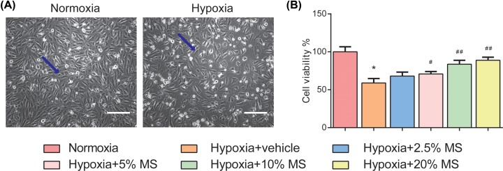
Figure 4. HSHS MS enhances endothelial cell viability after hypoxia in vitro.
(A) Representative images of normoxia and HUVECs after hypoxia (indicated by blue arrows). Scale bar = 300 μm. (B) The data of CCK8 cell viability assay are as follows. n=3. Data are presented as mean ± SD. *P<0.01 vs. Normoxia, #P<0.05 vs. Hypoxia+vehicle, ##P<0.01 vs. Hypoxia+vehicle.

