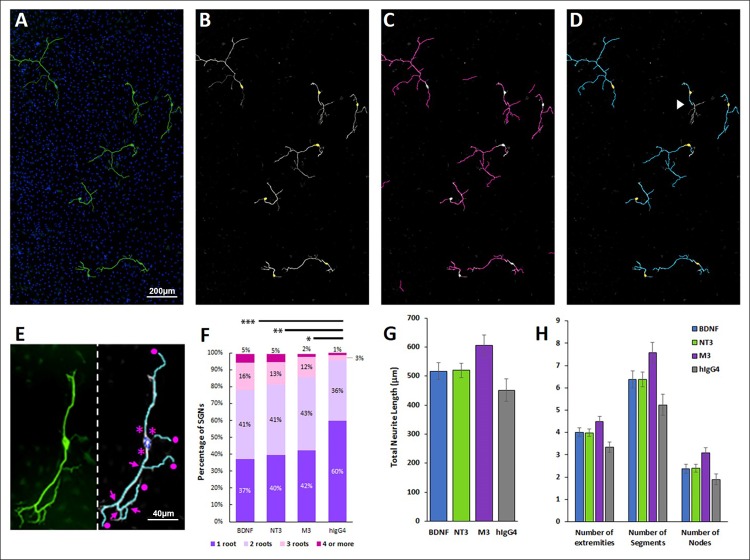
Fig 3. BDNF, NT-3, and M3 increase SGN complexity.
(A-D) SGNs were immunostained, imaged and batch analyzed using PerkinElmer Harmony 4.6. (A) SGNs were immunostained for neurofilament (green) and DAPI (blue) and imaged at 20x. All of the images in a single well were stitched together and analyzed. The analysis detects SGN somas (B, highlighted in yellow) and neurites (C, magenta). (D) Each neurite tree is assigned to a single cell for analysis. Depending on the stitching, occasionally neurites will not meet the criteria due to gaps that exceed a user-defined threshold (D, white arrowhead). (E) The number of roots (asterisk), extremities (circles), nodes (arrows), segments (discrete sections of neurite between nodes or extremities), and total neurite length were measured for each neuron. (F) Treatment with 10nM BDNF, 1nM NT-3, or 1nM M3 increased the number of roots compared to the IgG-treated control. (G-H) The total neurite length, as well as number of extremities, segments, and nodes per cell increased with BDNF, NT-3, and M3 treatments compared to hIgG4, but the differences were not statistically significant. *p< 0.05, **p<0.005, ***p<0.001. N = 67 to 834 neurons per treatment condition.

