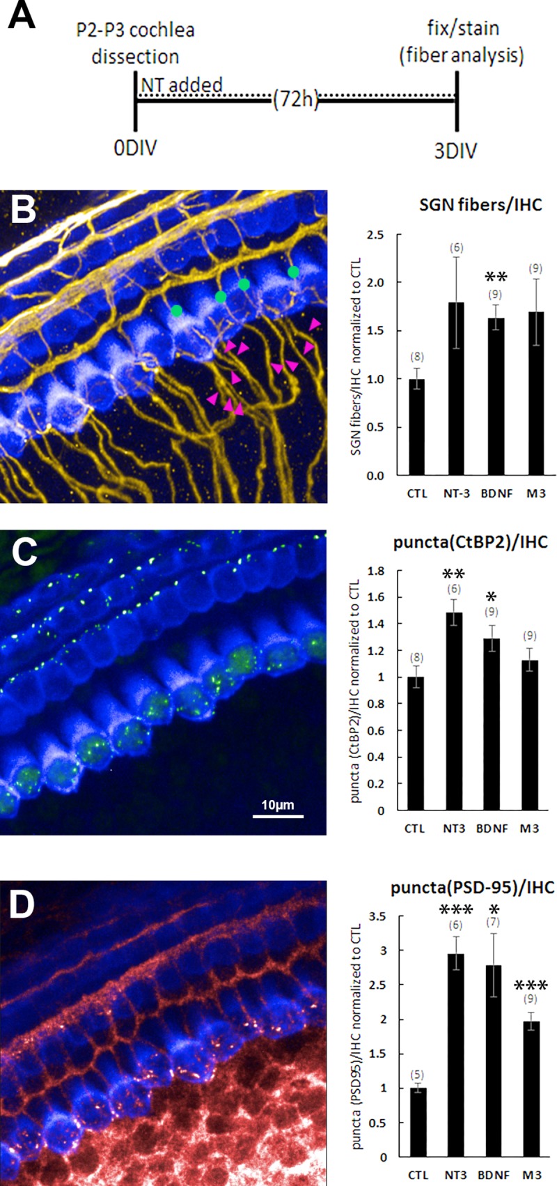
Fig 5. NT-3, BDNF and M3 promote formation of synapses and fiber growth in cochlear explants.

(A) Protocol for cochlea explantation and culture. (B) Explants were immunostained with Myo7A (blue) to identify hair cells and neurofilament (yellow) to label SGNs. The number of type I SGNs was calculated by counting all of the fibers approaching the hair cells and then subtracting those that continue to the outer hair cell layer. Green dots and magenta arrowheads represent a few examples of the fibers that were counted. Neurotrophins and M3 increased the number of type I SGN fibers contacting inner hair cells. (C) The pre-synaptic puncta were identified by an antibody that labels CtBP2 (green). Neurotrophins and possibly M3 increased the number of presynaptic puncta. (D) The postsynaptic puncta were identified by an antibody that labels PSD-95 (red). Neurotrophins and M3 increased the number of postsynaptic puncta. Bar graphs are normalized to control explants that were maintained in culture media but not treated with neurotrophic agents. The number of explanted tissues is indicated in parentheses and are combined from two (for PSD-95) or three (for SGNs and CtBP2) independent experiments. *p<0.05, **p<0.005, ***p<0.0005 (vs. CTL).
