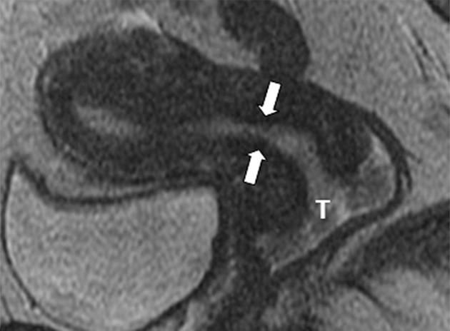
Figure 1a:

Detection of the tumor in the cervix and the location of the interval os. (a) Sagittal, (b) axial oblique at the internal os, (c) axial oblique at the external os, and (d) coronal oblique T2-weighted fast spin-echo images in a 28-year-old woman with stage IB1 endocervical adenosquamous carcinoma demonstrate predominantly exophytic intermediate T2-weighted SI tumor (T) adjacent to the low-SI cervical stroma (dashed arrow). The location of the internal cervical os (thick arrows) is defined as the entrance of the uterine vessels (thin arrows; best seen on axial oblique T2-weighted image) and narrowing of the uterine contour (best visualized on sagittal and coronal oblique T2-weighted images).
