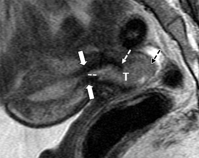
Figure 2a:

Tumor-to-internal os distance and presence of deep cervical stromal invasion. (a) Sagittal and (b) axial oblique T2-weighted fast spin-echo images in a 23-year-old woman with stage IB1 invasive adenocarcinoma of the uterine cervix demonstrate intermediate T2-weighted SI tumor (T) within 3 mm of the internal os. The internal os is seen as the narrowing of the uterine contour, where the low-SI fibrous cervical stroma changes to the intermediate-SI uterine myometrium (thick arrows; best seen on the sagittal T2-weighted image) and the entrance of the uterine vessels (thin arrows; best seen on axial oblique T2-weighed image). The tumor disrupts low-T2-SI inner cervical stroma (white dashed arrow) and extends up to the outer cervical stroma.
