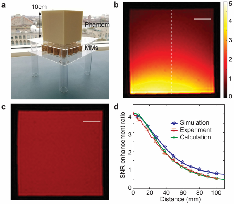
Fig. 5. Experimental results with and without metamaterials.
(a) Experimental setup of metamaterial array beneath 3D printed magnetic resonance imaging (MRI) phantom containing 2% agar gel. (b) MRI image in presence of 4 × 4 metamaterial array with unit cell diameter of 3 cm (transmission radio frequency (RF) power of MRI experiment reduced, see Methods; the dashed line used for comparison of signal-to-noise ratio (SNR) in (d)). (c) Control MRI image in the absence of metamaterial array (transmission RF energy not reduced, see Methods). Scale bars correspond to 2 cm in (b) and (c). (d) Comparison of SNR enhancement ratios of experimental, simulation, and calculation results. The bars for experimental results show the standard deviation of the measured SNR enhancement ratio.

