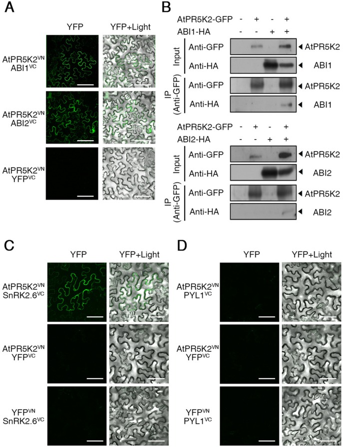
Figure 4.
AtPR5K2 interaction with ABI1, ABI2, and SnRK2.6 in vivo and in vitro. (A) Bimolecular fluorescence complementation (BiFC) analysis of AtPR5K2 and PP2Cs coexpressed in tobacco (Nicotiana benthamiana) leaves. VN and VC indicate the N- and C-terminal regions of Venus (eYFP), respectively. The epidermal cells were analyzed using confocal fluorescence microscopy and photographed after 48 h of incubation at 25°C. Scale bars represent 100 μm. (B) AtPR5K2 forms a complex with the PP2Cs. Each combination of 35S:AtPR5K2-GFP, 35S:ABI1-HA, and 35S:ABI2-HA were transiently expressed in tobacco plants. The proteins were immunoprecipitated with an alpha-green fluorescent protein (α-GFP) antibody and resolved with SDS-PAGE. The immunoblots were probed with an α-GFP antibody to detect AtPR5K2 or an α-HA antibody to detect ABI1 and ABI2. The minus (−) indicated empty vectors (35S:GFP or 35S:HA, respectively) as negative controls. (C and D) BiFC analysis of AtPR5K2 and SnRK2.6 (C) or PYL1 (D) coexpressed in tobacco leaves. The epidermal cells were analyzed using confocal fluorescence microscopy and photographed after 48 h of incubation at 25°C. Scale bars represent 100 μm.

