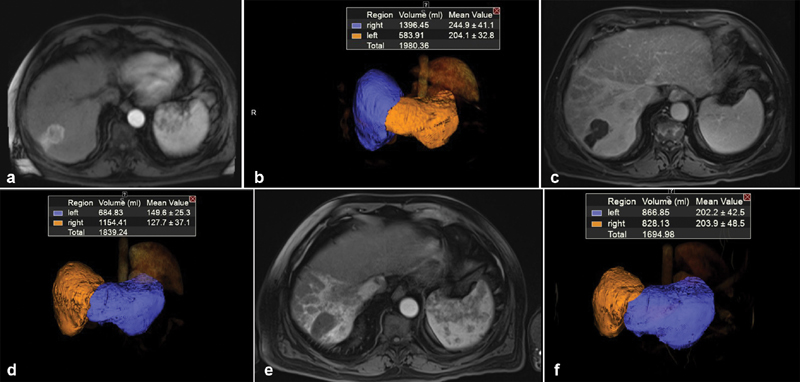
Fig. 2.

( a ) Baseline MRI of a 73-year-old male with solitary 3.5 HCC located in hepatic segment 7. ( b ) Volumetric assessment showing baseline FLR = 29.5%. ( c ) Three-month MRI follow-up post Y90 showing complete response of tumor by mRECIST criteria. ( d ) Volumetric assessment showing 3-month FLR = 37%. ( e ) Nine-month MRI follow-up post Y90 showing continuous complete response of tumor. ( f ) Volumetric assessment showing 9-month FLR = 51%.
