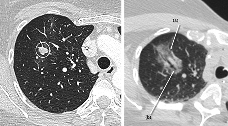
Fig. 3.

Conventional postablation appearance. Left: right upper lung lobe lesion prior to ablation. Right: the immediate postablation appearance of the same lesion, with (a) showing the expected ground-glass opacity encompassing the nodule with an hyperdense rim delimitating the ablation zone and (b) showing the ablation probe tract through the lesion. This typical appearance is easily noted in the 10-minute postablation scan.
