Fig. 5.

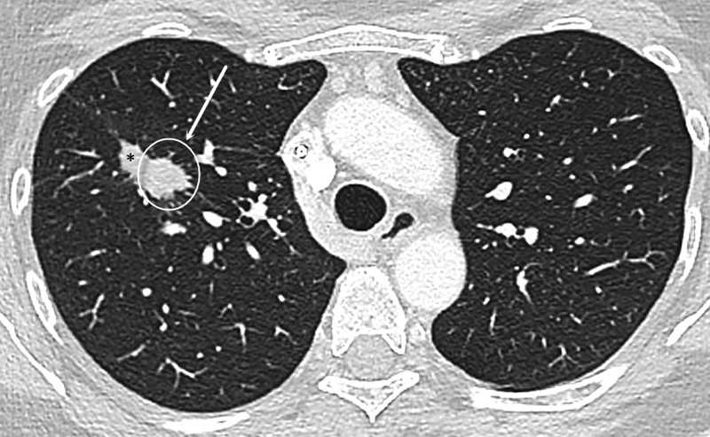
Example of recurrence. CT shows the ablated right upper lung lobe lesion at 6-month follow-up with a nodular contour of the anterior-medial aspect of the ablation zone (white circle). Note the involution of the adjacent ablation zone (*).

Example of recurrence. CT shows the ablated right upper lung lobe lesion at 6-month follow-up with a nodular contour of the anterior-medial aspect of the ablation zone (white circle). Note the involution of the adjacent ablation zone (*).