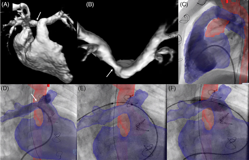
FIGURE 4.
XFM-guided right pulmonary artery (RPA) balloon angioplasty. Right pulmonary artery (RPA) balloon angioplasty performed in a patient with D-transposition of the great arteries s/p repair with LeCompte maneuver such that both pulmonary arteries course anterior to the ascending aorta. CMR data showed RPA stenosis with 40% of blood flow to the right lung and 60% to the left lung. (A) CMR delineation of the right-sided cardiac structures; (B) CMR delineation of the pulmonary artery bifurcation showing RPA stenosis (arrows); (C) right ventriculogram in the straight lateral projection; and (D) pulmonary artery angiogram in the RAO 30/CRAN 30 projection with XFM overlay of the right ventricle and pulmonary arteries in blue and the aorta in red; (E) balloon positioning demonstrating wire deformation of the RPA; and (F) proximal RPA balloon angioplasty

