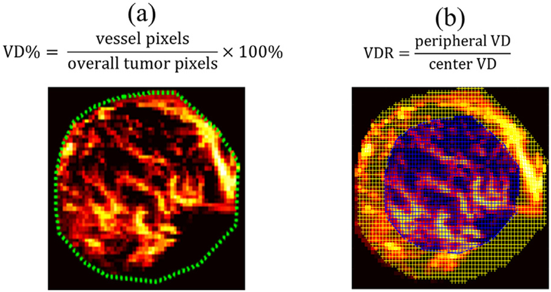
Fig. 1.
Vessel density (VD) and vessel density ratio (VDR) calculation using ultrasensitive ultrasound microvessel imaging (UMI). (a) VD calculation using an example UMI image of a 27-y-old woman with a fibroadenoma mass. The green dashed lines indicate the mass boundary. (b) VDR calculation example using the same mass. The blue-shaded area indicates the center region, and the yellow-shaded area indicates the periphery.

