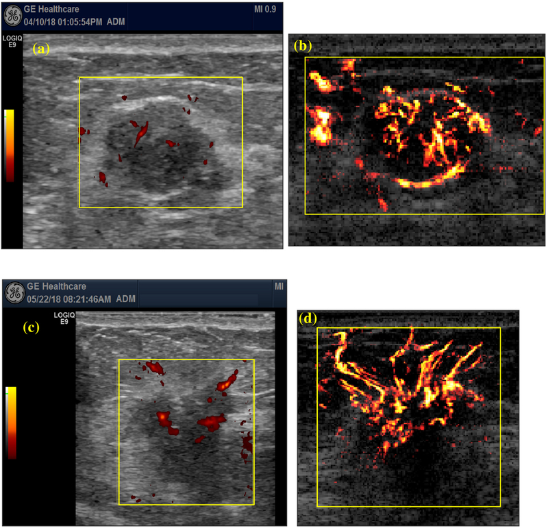
Fig. 2.
Comparison of vessel detection sensitivity between conventional Doppler and ultrasensitive ultrasound microvessel imaging (UMI). (a) Conventional Doppler and (b) UMI images of a 43-y-old woman with a fibroadenoma mass. Maximum mass diameter: 2.0 cm. (c) Conventional Doppler and (d) UMI images of a 40-y-old woman. Nottingham grade I (of III) invasive ductal. Maximum mass diameter: 1.9 cm.

