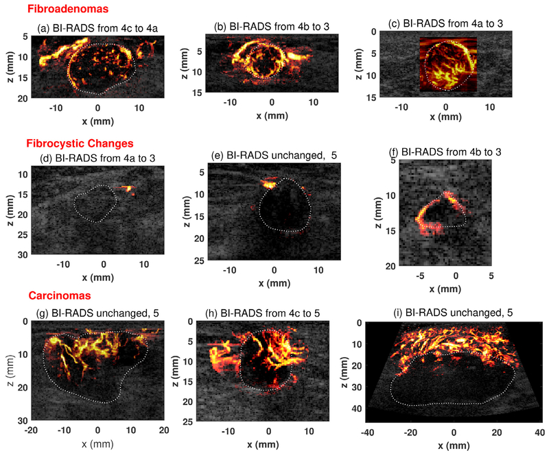
Fig. 4.
Representative ultrasensitive ultrasound microvessel imaging (UMI) of different types of breast masses. (a) Fibroadenoma from a 34-y-old woman. Maximum mass diameter: 2.34 cm. (b) Fibroadenoma from a 34-y-old woman. Maximum mass diameter: 1.3 cm. (c) Fibroadenoma from a 27-y-old woman; Maximum mass diameter: 2.0 cm. (d) A mass of fibrocystic change from a 67-y-old woman. Maximum mass diameter: 2.75 cm. (e) A mass of fibrocystic change from a 74-y-old woman. Maximum mass diameter: 1.3 cm. (f) A mass of fibrocystic change from a 52-y-old woman. Maximum mass diameter: 1.0 cm. (g) Nottingham grade II (of III) invasive lobular carcinoma from a 79-y-old woman. Maximum mass diameter: 3.0 cm. (h) Nottingham grade II (of III) invasive ductal carcinoma from an 83-y-old man. Maximum mass diameter: 1.7 cm. (i) Nottingham grade III (of III) adenocarcinoma (poorly differentiated) from a 43-y-old woman. Maximum mass diameter: 5.0 cm. The mass boundaries were delineated with white dashed lines to help readers appreciate the performance of UMI. Note that BI-RADS scoring and adjustment of BI-RADS categories using UMI images did not require delineation of tumor boundary. BI-RADS = Breast Imaging Reporting and Data System.

