Figure 2.
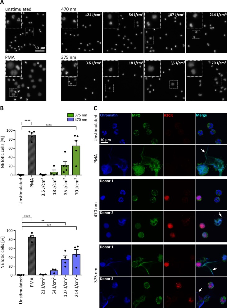
UVA and blue light induce the formation of NETs in a dose-dependent manner. (A) Representative fluorescence images of neutrophils exposed to different doses of LED-light [375 nm (3.5, 18, 35, and 70 J/cm2) or 470 nm (21, 54, 107, and 214 J/cm2)]. Decondensation of chromatin, stained by Hoechst, clearly increases with duration of light exposure. (B) NET rates significantly increase for both tested LEDs with light doses. Statistics: one-way-ANOVA with Bonferroni's multiple comparisons test (tested against unstimulated cells). **p < 0.01, ***p < 0.001, ****p < 0.0001. N = 3–5 independent experiments. Error bars = SEM. (C) Histone 3 becomes citrullinated (red/Alexa555) in early stages of chromatin decondensation (blue/Hoechst) after irradiation with both wavelengths. The decondensed chromatin colocalizes with MPO (green/alexa488) most prominently within the released NET fibers (arrows), similar to PMA-induced NETs. Confocal microscopy imaging of fixed samples. Cells were kept in RPMIcomp. + 10 mM HEPES.

