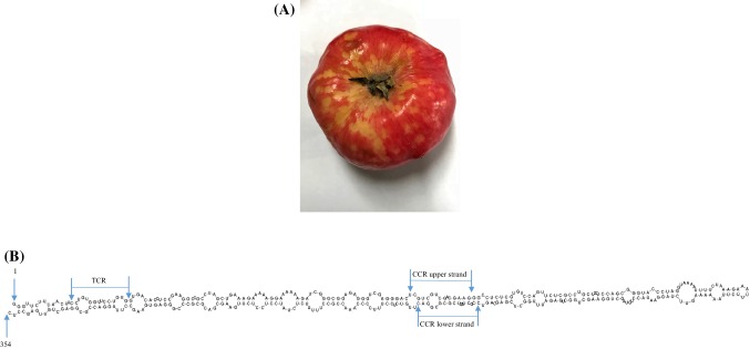
Fig. 1.
(A) Yellow and chlorotic fruit spots and peak-like symptoms on the skin of apples of cv. “Ilzer Rose” caused by ACFSVd. (B) Nucleotide sequence and proposed secondary structure of ACFSVd. Structures are represented for the lowest-energy form at 37 °C predicted using the model of Turner and Mathews [18], created using Geneious® software (Version 10.1.3). The first and the last nucleotide of ACFSVd, the TCR and the CCR lower and upper strands are indicated by arrows

