Fig. 1.

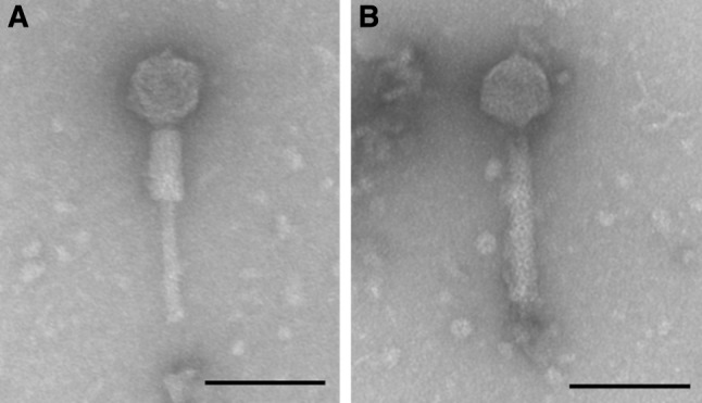
Transmission electron micrograph of bacteriophage R18C showing Myoviridae morphology with a contracted (A) and an uncontracted tail (B). Size measurements were performed on 15 intact individual phages. The bar represents 100 nm

Transmission electron micrograph of bacteriophage R18C showing Myoviridae morphology with a contracted (A) and an uncontracted tail (B). Size measurements were performed on 15 intact individual phages. The bar represents 100 nm