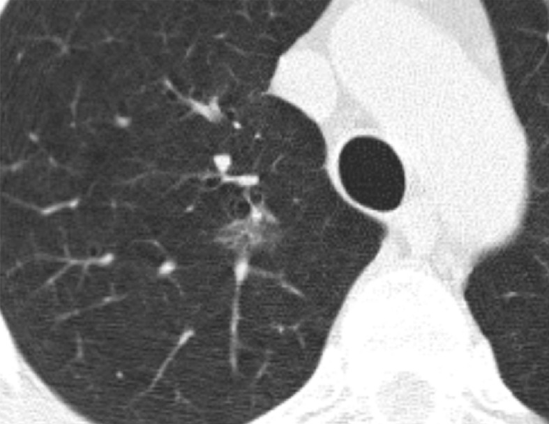
Figure 3b:
CT scans in a 57-year-old man undergoing low-dose lung cancer screening. (a) Baseline scan shows a right upper lobe pure ground-glass nodule measuring 11 mm (arrow). Nodule was classified as Lung-RADS category 2. (b, c) CT scans obtained at second annual follow-up show that this nodule grew to 15 mm and developed a 3-mm solid component (arrows). Malignancy was subsequently diagnosed.

