Fig. 5.

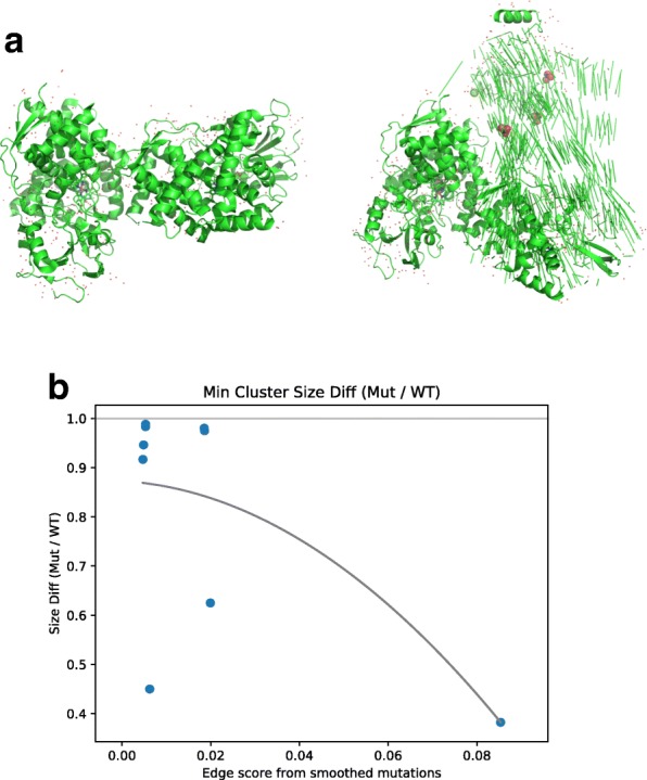
a Structure of prostaglandin I2 synthase, product of the PTGIS gene. Left: wild type, from PDB structure 2IAG, right: simulation of the impact of the high scoring edge mutation identified for this gene (amino acid substitution F293L). b Binding analysis of high and low scoring edges. For each edge we searched for protein structures for the two proteins connected by the edge in PDB. For pairs we found we simulated the impact of the mutation identified for that edge and used the ClusPro 2.0 docking tool to compare WT and mutated binding. Binding scores (y axis) represent ratio of maximum protein binding cluster with mutation vs. wild-type proteins. The lower the ratio the bigger the impact of the mutation. Curve is the best fit for a polynomial of degree 2. The curve indicates that as the edge score increases (x axis) the impact on binding increases as well
