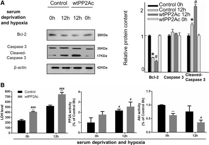
Fig. 7.
After PP2Ac overexpression, apoptosis and cell damage became more severe in L02 cells exposed to serum deprivation and hypoxia, so did Akt inactivation. L02 cells were transfected with RFP-wtPP2A plasmid (the wtPP2A group) or RFP-vector (the control group) for 48 h. Then cells were serum deprived and cultured under hypoxic conditions. a Left: representative images of western blots for apoptotic markers Bcl-2, caspase-3 and cleaved caspase 3. Right: semi-quantitative analysis with β-actin as an internal control. The results were normalized to the control 0 h group. b Left: LDH measurement; the unit: U/L. Middle and right: the PP2A or Akt activity measurement normalized to the control 0 h group. All data were expressed as the mean ± SEM. n = 3 for each group in each measurement. *p < 0.05 and **p < 0.01 versus control cells at the same time; #p < 0.05 and ###p < 0.001, 12 h versus 0 h, with the same transfection

