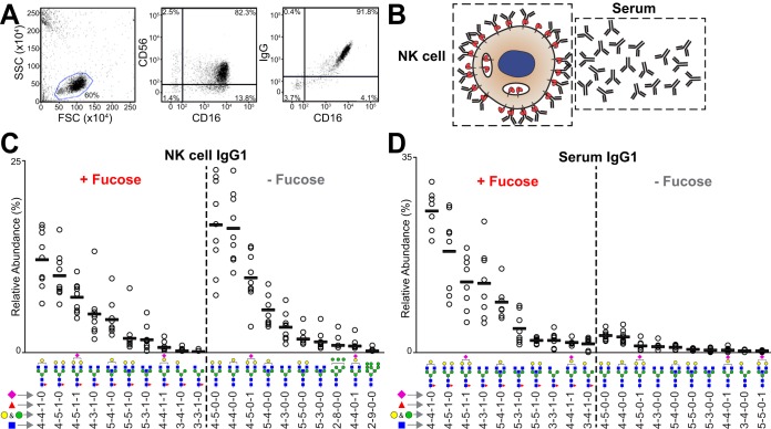
Fig. 7.
NK cells retain IgG1 lacking core fucose. A, Flow cytometry showing the collinearity of IgG and CD16a staining on primary human NK cells following cell isolation. B, Sources of IgG molecules (depicted as Y-shaped symbols). The top ten fucosylated and afucosylated IgG1 glycoforms isolated from primary NK cells (C) and serum (D). See also supplemental Tables S3 and S4.

