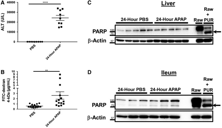
Figure 3.

Intestinal barrier deficits persist at 24 hours after APAP overdose despite a reduction in ileum PARP cleavage. Six‐ to eight‐week‐old male C57BL/6J mice were fasted for 12 hours and treated with 500 mg/kg APAP by IP injection. Food was returned to the animals, and they were restricted from water access starting from 8 hours after APAP injection. After an additional 12 hours, mice were gavaged with 1 g/kg 4‐kDa FITC–dextran. Water was then returned to the animals until they were euthanized 4 hours later (a total of 24 hours after APAP treatment). Serum was analyzed for levels of (A) ALT and (B) recovery of FITC–dextran. Data are represented as means ± SEM and n = 9‐15 mice per group from at least three independent experiments. Statistical analyses were made by the Student t test; **P < 0.01, ****P < 0.0001. Total protein lysates (25‐30 μg) of (C) liver and (D) distal ileum tissue were analyzed by western blot for PARP cleavage. Lysates of Raw 264.7 macrophages treated with or without 10 μg/mL PUR for 18 hours were included as controls. Arrows indicate cleaved PARP. Western blot images are representative of 10 to 15 mice per group from at least three independent experiments.
