
Figure 3.
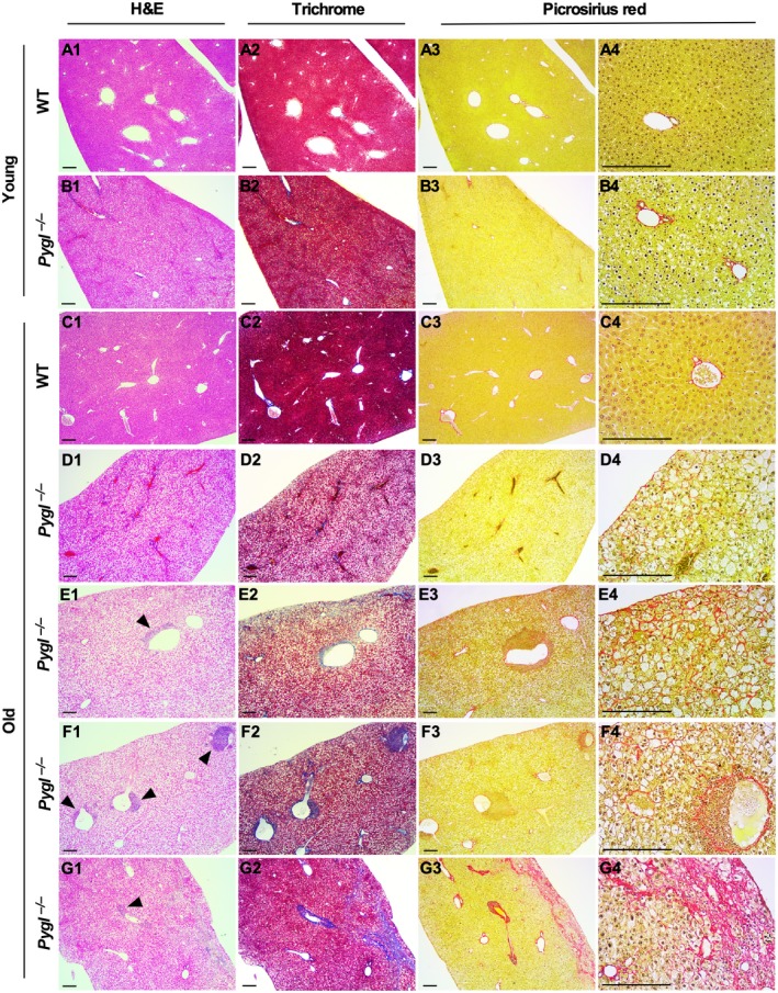
Histologic analysis of livers from WT and Pygl −/− mice. Livers from young WT (n = 6) and Pygl −/− (n = 9) mice and old WT (n = 7) and Pygl −/− (n = 13) mice were evaluated histologically by a veterinary pathologist. Representative images of liver sections stained with H&E, Masson's trichrome, and picrosirius red in young WT mice (A1‐A4), young Pygl −/− mice (B1‐B4), old WT mice (C1‐C4), and four individual old Pygl −/− mice (D1‐D4, E1‐E4, F1‐F4, G1‐G4). With picrosirius red staining, individual old Pygl −/− mouse exhibited distinct pathological patterns, including minimal collagen deposition in subcapsular areas (D3,D4); subcapsular and sinusoidal mild collagen deposition (E3,E4); minimal collagen deposition in perisinusoidal and periportal areas (F3,F4); and regionally severe fibrosis with central to central bridging and collapse of the intervening lobular structure (G3,G4). In picrosirius red staining, the images in the fourth column (A4‐G4) present higher magnification views of the images in the third column (A3‐F3). Scale bars represent 200 µm. Arrow heads indicate immune cell infiltrations.