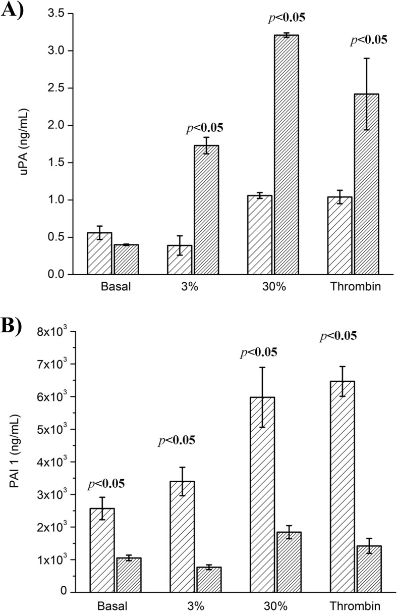
Fig. 2.

uPA and PAI 1 concentration under different stimulation conditions. The basal and thrombin condition consisted of HMEC-1 culture incubated with medium + 10% FBS and 1 nM thrombin and 2 mM CaCl2, respectively. The 3 and 30% fibrin γ/γ’ conditions consisted of HMEC-1 culture incubated with clots formed with purified fibrinogen (3% γ/γ’) and 30% fibrinogen γ/γ’ (by mixing commercial fibrinogen γ/γ and γ/γ’). A) uPA (ng/mL) and B) PAI (ng/mL) quantified by ELISA in the supernatant ( ) and cell lysate (
 ). The uPA concentration in the supernatant of basal condition was similar to that of cell lysate, while those of thrombin, 3 and 30% fibrin γ/γ’ were significantly higher in the cell lysate (p < 0.05). Instead, the PAI concentration was higher in the supernatant in all stimulatory and basal conditions (p < 0.05)
