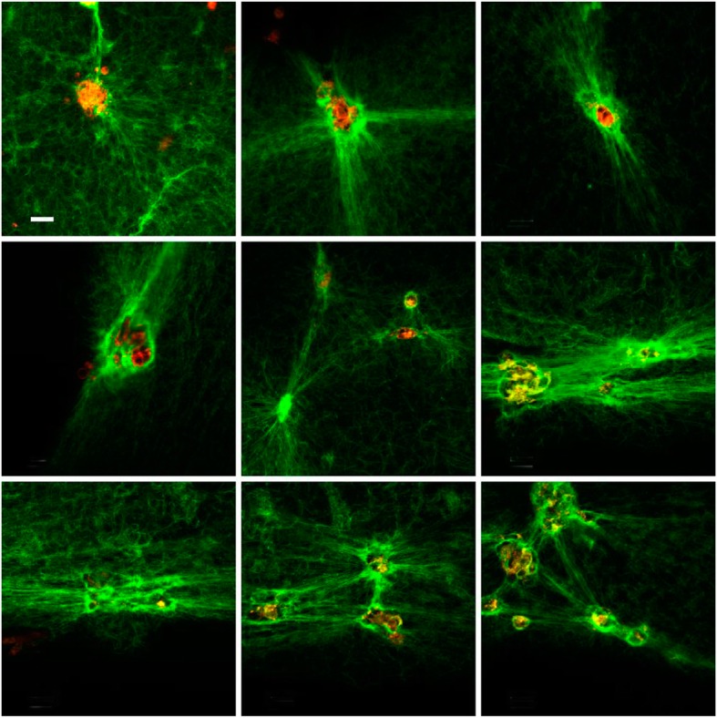
Fig. 5.

Fibrin 30% γ’ formed on the top of HMEC-1. Nine representative fields were selected at 10 μm from the cell surface. Fibrin fibers were labeled with Alexa 488-conjugated fibrinogen (green), and HMEC-1 cells with di-8-ANNEPS (red). Fibrin colocalizing with cell membrane appears yellow. The tool bar represents 30 μm
