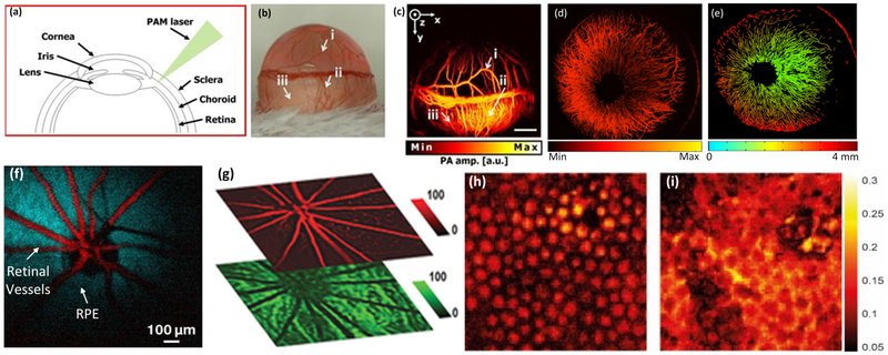
Figure 6.
Photoacoustic microscopy of the retinal: (a) structure of mouse eye, (b) a photograph of the mouse eye, (c) corresponding MIP PAM image of the corneal microvasculature in an albino mouse eye in (b), showing the iris, and limbal blood vessels as well as the retinal and choroidal vessels underlying the sclera [51]. (d) Photoacoustic microscopy of the iris vasculature of rat. (e) Depth-encoded PAM image [83]. (f) Pseudo-colored PAOM images of the retinal vessels and RPE [33]. (g) Segmented PAOM of the retinal and choroidal blood vessels. The retinal vessels are pseudo-colored in red, and the choroidal vasculatures are pseudo-colored in green [58]. (h,i) segmented PAM porcine retinal pigment endothelium (RPE) and choroid [84]. Adapted with permission from ref [33,51,83,84].

