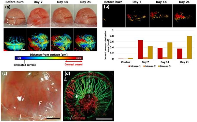
Figure 7.
PAOM of corneal neovascularization. (a) Surface-based depth encoded PAM images of corneal before and after alkali burn-induced corneal neovascularization at various times. (b) Segmented PAM image of supra-surface vessels and quantified corneal neovascularization area [51], (c) bright field microscopy image of the eye, and (d) PAM distinguishes between the iris (green) and corneal (red) vasculatures, due to their different depths. The green arrow points toward iris vessels; the red arrow points toward corneal neovascularization in the Z-slice. F: front, and L: limbus [85]. Adapted with permission from ref. [51,85].

