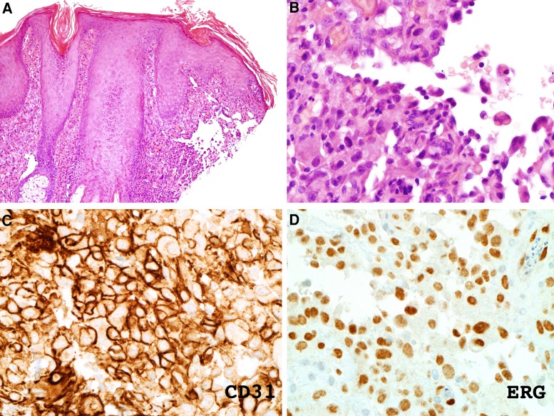
Figure 2.
(A,B) Hematoxylin and eosin staining of skin biopsy shows dermal infiltrate of pale eosinophilic cells that have characteristic features of “dissecting the collagen bundles” and atypical, enlarged, and hyperchromatic nuclei. (C,D) Immunostaining shows staining for vascular markers CD31 and ERG (labeled).

