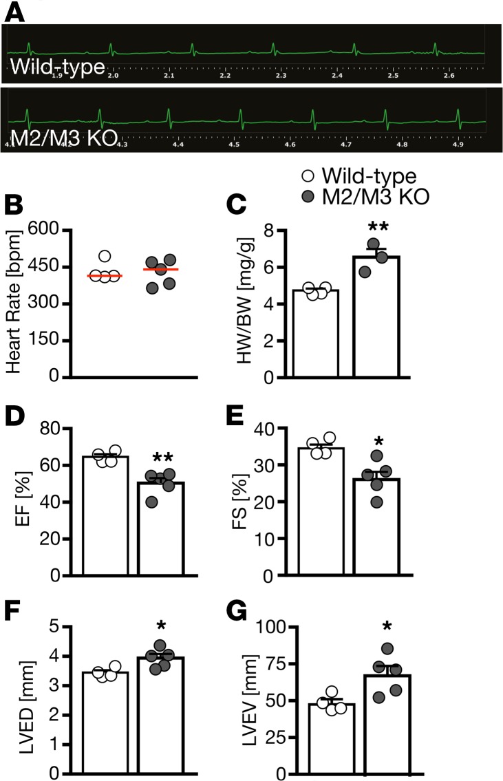
Figure 4. Mice lacking both M2 and M3 muscarinic receptors show impaired cardiac performance.
(A) Representative echocardiograms of WT and M2/M3 double-KO mice reveal no abnormalities in cardiac rhythm. n = 4–5 mice per genotype (5–6 months of age, mixed sex). (B) Heart rate was not altered in M2/M3 double-KO mice. n = 4–5 mice per genotype. (C) M2/M3 double-KO mice had a higher heart weight (HW) to body weight (BW) ratio. This was due to an overall reduced size of M2/M3 double-KO mice. **P = 0.0057, 2-tailed t test. n = 4–5 mice per genotype. (D) M2/M3 double-KO mice had a reduced ejection fraction (EF). **P = 0.0040, 2-tailed t test. n = 4–5 mice per genotype. (E) Fractional shortening (FS) was also reduced in M2/M3 double-KO mice. *P = 0.0129, 2-tailed t test. n = 4–5 mice per genotype. (F) At the same time, left ventricular end-diastolic diameter (LVED) was increased in M2/M3 double-KO mice. *P = 0.0272, 2-tailed t test. n = 4–5 mice per genotype. (G) M2/M3 double-KO mice further displayed a larger left ventricular end-diastolic volume (LVEV). *P = 0.0303, 2-tailed t test. n = 4–5 mice per genotype. Bar graphs show mean ± SEM and circles indicate individual mice.

