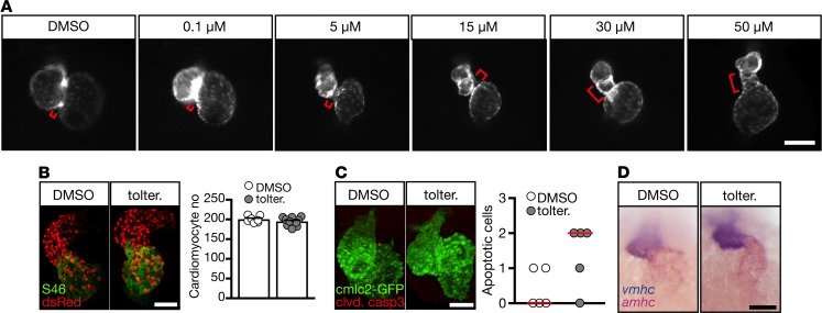
Figure 7. Tolterodine treatment causes elongation of the AVC.
(A) Whole-mount live images of fluorescent zebrafish hearts [Tg(myl7:EGFP)] at 48 hpf. The AVC (indicated by a red bracket) is elongated upon tolterodine treatment. (B) Cardiomyocyte number remained unaltered in tolterodine-treated zebrafish. Images show confocal stacks of Tg(-5.1myl7:DsRed2-NLS) zebrafish. DsRed-expressing nuclei of cardiomyocytes were enhanced by staining with an anti-DsRed antibody. Atria were visualized by staining of atrial myosin (S46). Graph shows individual embryos as circles and mean ± SEM. n = 7–10 embryos. (C) Tolterodine did not induce apoptosis in cardiomyocytes as shown by the lack of cleaved caspase-3–positive cardiomyocytes. Images show confocal stacks of Tg(myl7:EGFP) hearts. Circles show individual embryos. P = 0.1429, 2-tailed Mann-Whitney U test. n = 5 embryos each. (D) Chamber specification was not affected by tolterodine. Atrial (amhc) and ventricular (vmhc) myosin visualized by double in situ hybridization. n = 3 experiments with 34–50 embryos. Scale bars: 100 μm (A), 50 μm (B–D).

