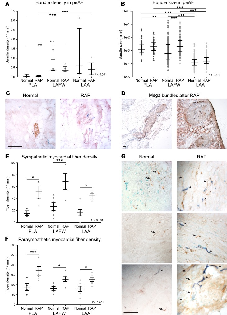
Figure 1. Persistent AF induced by rapid atrial pacing (RAP) causes a global increase in left atrial autonomic nervous fibers.
(A) Nerve bundle density in the posterior left atrium (PLA), left atrial free wall (LAFW), and left atrial appendage (LAA) of normal dogs and animals subjected to RAP is shown as median with interquartile range. n = 3, 4, 5, 5, 4, and 6. (B) Combined nerve bundle size in the PLA, LAFW, and LAA of normal dogs and animals subjected to RAP is shown as median with interquartile range. n = 53 and N = 4 for normal PLA, n = 69 and N = 6 for RAP PLA, n = 138 and N = 6 for normal LAFW, n = 125 and N = 6 for RAP LAFW, n = 112 and N = 5 for normal LAA, and n = 160 and N = 6 for RAP LAA. (C) Representative micrographs of nerve bundles in left atrial tissue of normal dogs (left) and after RAP (right) subjected to IHC for AChE (brown) and DBH (blue). (D) Examples of very large bundles seen in the PLA and LAFW after RAP. For C and D, scale bar: 500μm. (E) Sympathetic and (F) parasympathetic myocardial fiber density in the PLA, LAFW, and LAA of normal dogs or after RAP is shown mean ± SEM. For sympathetic fibers, n = 3, 4, 6, 4, 6, and 4. For parasympathetic fibers, n = 4, 6, 5, 6, 6, and 7. (G) Representative micrographs of IHC for AChE (brown) and DBH (blue) on left atrial tissue of normal dogs (left) and after RAP (right). Examples of parasympathetic (AChE+) fibers indicated with arrows. Examples of sympathetic (DBH+) fibers indicated with arrowheads. Scale bar: 250 μm. Two-way ANOVA significance indicated in graphs, after log transformation for nonparametric values. *P < 0.05; **P < 0.01; ***P < 0.001 for pairwise comparison with Holm-Sidak method.

