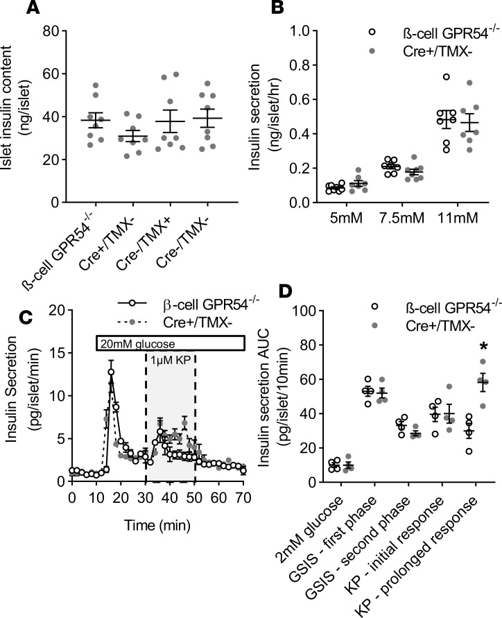
Figure 3. Effects of β cell GPR54 deletion on islet function in vitro.
(A) There were no significant differences in islet insulin content between β cell GPR54–/– mice and any control groups (Cre+/TMX–, Cre–/TMX+, and Cre–/TMX–). Mean ± SEM, n = 9. (B) In static incubation experiments there was no significant difference in the insulin secretory response to physiological glucose concentrations between β cell GPR54–/– and Cre+/TMX– islets. (C) In perifusion experiments there was no significant difference in basal insulin secretion at 2 mM glucose or first- or second-phase insulin secretion in response to 20 mM glucose in β cell GPR54–/– compared to Cre+/TMX– islets. Exposure of Cre+/TMX– islets to kisspeptin (1 μM, 30–50 minutes) resulted in a sustained enhancement of second-phase insulin secretion for the duration of kisspeptin administration. However, in β cell GPR54–/– islets this response was transient and not maintained beyond 10 minutes as determined by comparison of individual time points and (D) AUC over 10-minute phases of the perifusion (2-tailed Student’s t test; KP — prolonged response; P = 0.007). Mean ± SEM; n = 8 (A); n = 7–8 (B); n = 4 (C and D); data representative of experiments conducted 2–3 times; *P < 0.05.

