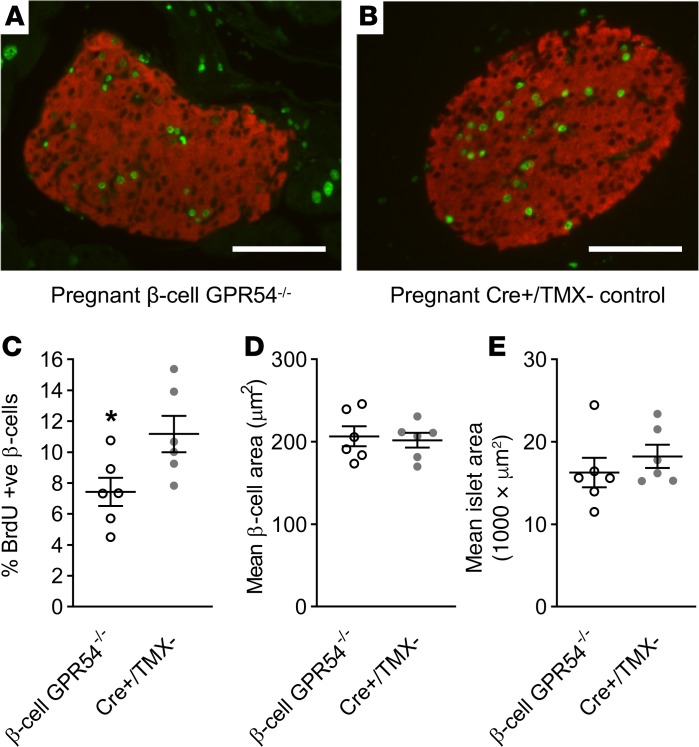
Figure 5. Effects of deletion of β cell GPR54 on pregnant β cell mass.
Representative illustrative images of immunostaining for the measurement of β cell proliferation in pregnant (A) β cell GPR54–/– and (B) Cre+/TMX– islets showing merged BrdU staining (shown in green) and insulin staining (shown in red). Scale bars: 100 μm. (C) β cell GPR54–/– mice administered BrdU from days 10–18 of pregnancy had significantly reduced levels of BrdU labeling compared with pregnant Cre+/TMX– mice (2-tailed Student’s t test, P = 0.041). At day 18 of pregnancy there was no significant difference in either (D) mean overall islet area or (E) size of individual β cells between β cell GPR54–/– and Cre+/TMX– mice. Mean ± SEM; n = 6 (C–E); *P < 0.05.

