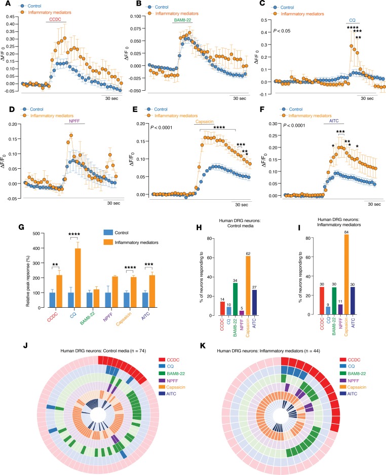
Figure 12. Human DRG neurons respond to pruritogenic agonists for TGR5, in addition to the MrgprX1 agonists chloroquine and BAM8–22.
(A–F) Human DRG neurons were cultured in control media, and in order to simulate a pathological state, a subset of cultures incubated with inflammatory mediators. This consisted of histamine (10 μM), PGE II (10 μM), serotonin (10 μM), and bradykinin (10 μM) being incubated with the neurons for 2 hours before Ca2+ imaging experiments commenced. Human DRG neurons from this cohort are referred to as inflammatory mediators. Grouped data of Ca2+ responses in control (n = 74) and inflammatory mediator (n = 44) cultured human DRG neurons to application of the (A) TGR5 agonist CCDC (100 µM), MrgprX1 agonists (B) BAM8-22 (2 μM), (C) CQ (1 μM), (D) NPFF (2 μM), (E) TRPV1 agonist capsaicin (100 nM), and (F) TRPA1 agonist AITC (50 M). Two-way ANOVA indicate responses to CQ (*P < 0.05), capsaicin (***P < 0.001), and AITC (**P < 0.01) are all significantly increased in neurons that had been exposed to inflammatory mediators. (G) Peak response of neurons to CCDC (**P < 0.01), CQ (****P < 0.0001), capsaicin (****P < 0.0001), and AITC (***P < 0.001) were all significantly increased in human DRG neurons incubated with inflammatory mediators. (H and I) Human DRG neurons from (H) control and (I) inflammatory mediator cultures responding to CCDC, CQ, BAM8-22, NPFF, capsaicin, and AITC. (J and K) Donut plot analysis showing the functional coexpression profiles as determined by Ca2+ imaging of (J) 74 individual human DRG neurons from control cultures and (K) 32 individual human DRG neurons from inflammatory mediator cultures in response to CCDC, CQ, BAM8-22, NPFF, capsaicin, and AITC. Data presented are mean ± SEM. P values determined by 2-way ANOVA and Bonferroni post hoc tests (significance indicated within panels) (A–F) or unpaired t tests (G).

