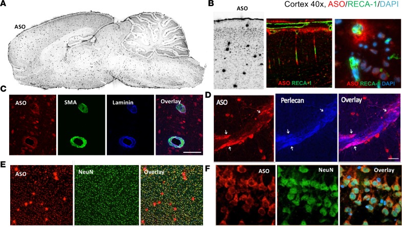
Figure 7. Perivascular and neuronal association of ASO 24 hours after dosing.
(A) Punctate perivascular staining of ASOs by IHC throughout brain at 24 hours after IT dosing. (B) Colocalization of ASOs and the vascular endothelial marker Reca1 in cerebral cortex (×10 magnification left 2 images, ×40 magnification right image). (C) Colocalization of Cy7-ASO with vascular α-smooth muscle actin (αSMA) and basement membrane component laminin α2 (×20 magnification). (D) Colocalization of ASOs with basement membrane component perlecan (×20 magnification). (E) ASO colocalization with neurons and vessels in cerebral cortex (×10 magnification). (F) ASO colocalization with neuronal cells in hippocampus (×40 magnification).

